Authors:
Anusree P.S.、Bikram Keshari Parida、Seong Yong Moon、Wonsang You
Paper:
https://arxiv.org/abs/2408.09358
Introduction
In the realm of dental health care, imaging modalities such as Cone Beam Computed Tomography (CBCT) and Panoramic X-rays are indispensable tools. CBCT provides three-dimensional views of a patient’s head, enhancing diagnostic capabilities, while Panoramic X-rays capture the entire maxillofacial region in a single image with minimal radiation exposure. However, the need for both imaging techniques can lead to increased radiation exposure and patient discomfort. This study introduces a novel method to synthesize Panoramic X-rays from existing CBCT data, thereby eliminating the need for additional scans and reducing radiation exposure.
Related Work
Existing Methods and Challenges
Current methods for synthesizing Panoramic X-rays from CBCT data involve delineating an approximate dental arch and creating orthogonal projections along this arch. However, these methods face several challenges:
– Lack of Standardization: No golden standard exists for dental arch extraction, affecting the quality of synthesized X-rays.
– Patient-Specific Variability: Existing methods struggle with patients having missing or nonexistent teeth and those with severe metal implants.
– Manual Intervention: Accurate identification of craniofacial landmarks often requires expert intervention.
Previous Approaches
Several approaches have been proposed to address these challenges:
– Curve Fitting Algorithms: Automated techniques using polynomial or Bezier curve fitting to approximate dental arches.
– Dynamic Rotation Centers: Contemporary panoramic systems use movable rotation centers to trace an elliptical path during jaw scanning.
– Heuristic Methods: Park et al. defined a quadratic curve for dynamic rotation trajectories, while Kwon et al. isolated panoramic projection data from CBCT projection data.
Research Methodology
Proposed Method
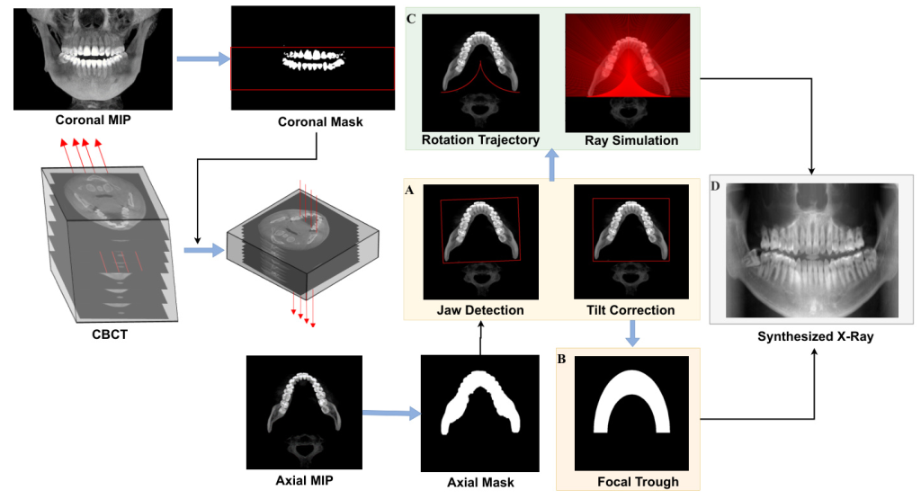
The proposed method synthesizes Panoramic X-rays from CBCT data using a simulated projection geometry with dynamic rotation centers. The methodology involves:
1. Jaw Detection and Tilt Correction: Detecting the jaw position and correcting any misalignment due to head tilt.
2. Focal Trough Definition: Defining an elliptical focal trough encompassing the maxilla and mandible.
3. Trajectory Formulation: Simulating the movement of the source-detector system along a semi-elliptical trajectory.
4. X-ray Projection Generation: Generating multiple X-ray projections along the trajectory and summing them to produce the final Panoramic X-ray image.

Jaw Detection and Tilt Correction
The process begins with detecting the jaw position using Coronal and Axial Maximum Intensity Projections (MIPs) from the CBCT data. Misalignments are corrected by adjusting the window level to enhance contrast and eliminate extraneous tissues. Contour detection algorithms are then used to extract the jaw shape.
Focal Trough Estimation
Elliptical focal troughs are defined to encompass the entire mandible and maxilla, with thicker regions near the incisors and thinner regions near the molars. This approach minimizes unwanted anatomical structures and ghost images in the X-ray output.
Panoramic Scan Simulation
An elliptical trajectory model is developed to simulate the movement of the source-detector unit. Rays are traced along this trajectory, and points along these rays are sampled and aggregated into single pixels using Beer Lambert’s equation.
X-ray Synthesis
The synthesized X-ray images are generated by applying Beer Lambert’s absorption-only attenuation law to the sampled points along the rays. CBCT windowing techniques are used to enhance the visibility of incisors and other critical structures.
Experimental Design
Data Collection
The study utilized clinical dental head CBCT scans of 600 patients from the Chosun School of Dentistry in Gwangju, South Korea. The dataset included 16-bit grayscale images with resolutions ranging from 0.25 to 0.5 mm, captured using the CS900 scanner from Carestream Health and the Planmeca VISOG7 scanners. Real panoramic X-rays corresponding to each patient were also available for quality assessment.
Validation and Comparison
The proposed algorithm was validated across the dataset, demonstrating robust outcomes in jaw detection and tilt correction. The synthesized panoramic X-rays were compared against two baseline models using qualitative and quantitative metrics.

Results and Analysis
Qualitative Comparison
The synthesized panoramic X-rays were compared with real X-rays and those generated by baseline models. The proposed method showed superior performance in accurately representing anatomical structures, particularly near the incisors, and minimizing distortions and ghost images.

Quantitative Evaluation
The efficacy of the proposed method was evaluated using Learned Perceptual Image Patch Similarity (LPIPS) and Structural Similarity (SSIM) metrics. The results indicated that the proposed method outperformed the baseline models in producing higher-quality panoramic X-ray images.

CBCT Preprocessing
CBCT windowing techniques were strategically employed to enhance the quality of input data. Maximum Intensity Projections (MIP) were used to identify the Region of Interest (ROI) containing the teeth and jaw, facilitating accurate jaw detection and tilt correction.

Conventional vs. Synthesized X-rays
The synthesized X-rays demonstrated comparable diagnostic utility to conventional panoramic X-rays, capturing intricate details such as dental fixtures, bone loss, and anatomical structures with high fidelity.

Limitations
Despite its advantages, the proposed method has limitations, including the need for dynamic adjustment of the focal trough to conform to the patient’s jaw shape and the potential impact of scattering from metal implants and other artifacts.

Overall Conclusion
This study presents a novel approach for synthesizing Panoramic X-rays from CBCT data using simulated projection geometry with dynamic rotation centers. The proposed method addresses several challenges associated with existing techniques, demonstrating robust performance across a diverse dataset. The findings highlight the potential of this approach to enhance diagnostic capabilities while reducing radiation exposure and patient discomfort.

Acknowledgments
The authors thank Prof. Bumshik Lee and Prof. Sungyong Moon from Chosun University for their technical discussions and data collection. They also thank Youngchan Lee and Gyubin Lee from AIIP Lab, Sun Moon University for their support.
Funding
This work was supported by the Basic Science Research Program (NRF-2022R1F1A1075204), the BK21 FOUR, and the Regional Innovation Strategy Project (2021RIS-004) through the National Research Foundation of Korea (NRF) funded by the Ministry of Education, South Korea.
Data Availability
The datasets used in this study are not publicly available but can be requested from the corresponding author.
Correspondence
Correspondence and requests for materials or datasets should be addressed to W. You.

